Neurosurgery Chair Invited to Speak to Congressional Neuroscience Caucus May 10

Keith L. Black, MD, will describe an experimental, noninvasive method to diagnose Alzheimer’s disease before cognitive symptoms begin Los Angeles – May 8, 2013 – Keith L. Black, MD, chair of the Department of Neurosurgery at Cedars-Sinai Medical Center, will present information to the Congressional Neuroscience Caucus on a new, experimental method to provide early detection of Alzheimer’s disease. The noninvasive diagnostic procedure — which is in clinical trials — looks at the retina of the eye for early changes signaling the onset of Alzheimer’s. Black, director of the Maxine Dunitz Neurosurgical Institute, director of the Johnnie L. Cochran, Jr. Brain Tumor Center and the Ruth and Lawrence Harvey Chair in Neuroscience at Cedars-Sinai, is one of 10 speakers invited to participate at the panel’s second annual session on brain mapping, which will be from 10 a.m. to noon EST May 10 in the Gold Room of the Rayburn House Office Building, 45 Independence Ave. SW, Washington, D.C. The Congressional Neuroscience Caucus, co-chaired by Reps. Earl Blumenauer and Cathy McMorris Rodgers, aims “to build awareness of the intrinsic role brain research plays in understanding ourselves and our society, to help communicate the progress and the benefits of this research, and to help inform federal policy,” according to Blumenauer’s website. Members embrace “policies that encourage support for funding of quality neuroscience research (and) that enhance translation and dissemination of discoveries to maximize the medical and societal benefits of research.” Cedars-Sinai researchers showed that beta-amyloid protein plaques associated with Alzheimer’s disease occur not only in the brain but also in the retina in the back of the eye; the deposits can be seen in the retina even before they begin to accumulate in the brain. With these advances, the researchers developed a device that enables doctors to look through the eye — just as an ophthalmologist diagnoses retinal disease — to visualize amyloid plaques in the retina. Results of preclinical studies were encouraging, and the researchers now are working with major pharmaceutical companies to evaluate the technology in patient trials. The objective is to provide a quick, inexpensive, noninvasive way to screen patients for Alzheimer’s in its first stages so emerging treatments may be started early to slow or stop the progression of the disease. The Congressional Neuroscience Caucus’s annual brain mapping program is presented with the support of the Brain Mapping Foundation.
Society for Brain Mapping and Therapeutics (SBMT) announce the formation of American Board of Brain Mapping, its 2013 award recipients and its Brain Mapping Day at the US Congress
LOS ANGELES, April 12, 2013 /PRNewswire-USNewswire/ — President Obama said in his April 2nd speech “We have been a nation of dreamers and risk-takers; people who see what nobody else sees sooner than anybody else sees it. We do innovation better than anybody else — and that makes our economy stronger. When we invest in the best ideas before anybody else does, our businesses and our workers can make the best products and deliver the best services before anybody else.” Babak Kateb, Founding Chairman of the Board of SBMT, attended the White House event at which President Obama announced a new federal effort to fund research that will constitute a major new Brain Mapping initiative. The SBMT Board of Directors had previously submitted a whitepaper to the White House and is part of the national dialogue about President Obama’s Brain Mapping Initiative. “SBMT and Brain Mapping Foundation are naturally well positioned in playing a significant role in President Obama’s initiative. We are planning to establish a NanoBioElectronic Consortium, which will focus on integrating nanotechnology, stem cell, cellular and molecular biology, immunology, device and imaging,” said Babak Kateb, President of the Brain Mapping Foundation, Director of National Center for NanoBioElectronics, and Editor of the Textbook of Nanoneuroscience and Nanoneurosurgery. “We are also establishing the American Board of Brain Mapping in order to certify specialists in this field and planning to have a cross disciplinary doctoral program in nanobioelectronics in collaboration with one of the most prestigious universities in the US. This will help us integrate and translate technologies across multiple disciplines into neuroscience and train new generations of cross disciplinary thinkers and scientists.” The BRAIN Initiative is going to fund research on cutting-edge technologies, which could probe and or image the brain in order to better understand its structure and neuropatho-physiology as well as breaking new ground on the treatment, cure and prevention of neurological disorders such as Alzheimer’s disease, epilepsy, brain cancers and neurotrauma. “The standardization of training, technologies and introduction of technological solutions for neurological disorders need a body that could play a neutral role. SBMT for sure has the history, scientific credibility and the credential to ensure President Obama’s Initiative is a success,” said Warren Grundfest, Member of the board of SBMT, Professor Department of Bioengineering, Department of Electrical Engineering, Department of Surgery, UCLA Geffen school of Medicine The whitepaper, which is submitted by the SBMT board of directors to the White House, NIH, DARPA and DoD, calls for the formation of the National Alliance for NanoBioElectronics (NANBE), National Network for Human Brain and Specimen Banks (NNHBSB) and National Data Repository and Analysis for Neuroscience (NDRAN). “We examined the entire national policy in neuroscience and tried to come up with a truly unique approach in order to address some of the fundamental problems in the field; we also believe that board certification is a necessary step toward standardization of the science and the technology in the field of brain mapping,” said Gary Steinberg, member of the SBMT board, Bernard and Ronni Lacroute-William Randolph Hearst Professor of Neurosurgery and the Neurosciences Director, Stanford Institute for Neuro-Innovation and Translational Neurosciences Chairman, Department of Neurosurgery, Stanford University School of Medicine SBMT is holding its annual meeting at the Baltimore Convention Center on May 12-14, featuring more than 45 scientific sessions and nearly 300 invited speakers. The meeting will cover all aspects of Brain and spinal cord mapping including neurosurgery, radiology, neurology, neuroscience, stem cell, engineering, psychiatry, immunology, brain bank, military medicine, space medicine and neurophtolmology, neurodegenerative disorders, autism, Parkinson’s disease and radiation oncology. This program is also jointly sponsored by the International Society for Magnetic Resonance in Imaging (ISMRM) this year and in 2012 was jointly sponsored by American Association of Neurological Surgeons (AANS). “SBMT is the most interdisciplinary and collaborative organization in the world in the field of Brain Mapping; the organization has brought together leading industry, academics, government entities, international partners and non-profits in order to introduce game-changing technologies into the field and in order to better prevent, diagnose and treat neurological disorders,” said Mitchel Berger, President of AANS, member of the board of SBMT, Professor and Chairman, Department of Neurological Surgery, Kathleen M. Plant Distinguished Professor, Director, Brain Tumor Surgery Program, Director, Neurosurgical Research Centers, Brain Tumor Research Center Pioneer in Medicine awards will be presented by the SBMT to Drs.: Congressman Jim Moran (D), Earl Blumenauer (D) and Cathy McMorris-Rodgers(R) will be accepting the Pioneer in Healthcare Policy Award for their role in advocating policies, which have been positively impacting neuroscience. Humanitarian award of Brain Mapping Foundation goes to the Founder of Cogent Inc., Ming Hsieh for his significant role in pushing the boundaries of the science through USC Ming Hsieh institute. Eric Bailey, Founder and CEO of Neurologica and Reese S. Terry Jr, co-founder of Cyberonics Inc are the recipients of the Pioneer in technology development. Brain Tumor Survivor, Singer/Songwriter, Beth Nielsen Chapman will be accepting the Beacon of Courage and Dedication award from the Foundation. “We are truly pleased to have such an amazing group of scientists, a key policymaker and industry leaders as well a talented brain tumor survivor and a humanitarian on our award list,” said Michael Roy, President of SBMT (2012-13), Professor of Medicine and Director of Internal Medicine at Uniformed Services University Health Science. SBMT is also holding a Brain Mapping Day at the US Congress on May 10th 2013 from 10-12 noon at the Gold Room in Reyburn building. “This is an annual event, which is focused on educating policymakers about the state-of-the-art brain mapping and therapeutics,” said Sujit S. Prabhu, member of the board of SBMT, Associate Professor, Department of Neurosurgery, MD Anderson Cancer Center. About Society for Brain Mapping and Therapeutics:www.WorldBrainMapping.org About Brain Mapping Foundation:www.BrainMappingFoundation.org Media Contact:Press@WorldBrainMapping.orgOrLeo Balthazor Leo@WorldBrainMapping.Org 310-5006196 SOURCE Society for Brain Mapping and Therapeutics
Brain Activity Map Project in Planning Stages
The US government is in the planning stages of a massive project to map the activity of the human brain, Story C. Landis, PhD, director of the National Institute of Neurological Disorders and Stroke, confirmed in an interview with Medscape Medical News this week. President Barack Obama alluded to investing in the “Brain Activity Map” project in his State of the Union address, saying, “Every dollar we invested to map the human genome returned $140 to our economy. Today, our scientists are mapping the human brain to unlock the answers to Alzheimer’s; developing drugs to regenerate damaged organs; devising new material to make batteries 10 times more powerful. “Now is not the time to gut these job-creating investments in science and innovation,” he said. “Now is the time to reach a level of research and development not seen since the height of the Space Race. We need to make those investments.” Brain Activity Map Project A “concrete plan” for exactly how the project will unfold has yet to be ironed out, Dr. Landis said. It’s also not clear yet how much money the federal government will provide for the project, but a ballpark figure discussed is $300 million a year for a project slated to last 10 years. Dr. Landis said “work is now ongoing putting together a consortium of federal agencies that would be participating and there is interest from a number of different private foundations.” The Brain Activity Map project will piggyback on the Human Connectome Project, the National Institutes of Health–funded project to create a high-resolution map of the major structural and functional connections in the human brain. There has been “extraordinary progress” in understanding aspects of human brain organization, Dr. Landis said. “The Human Connectome Project is now beginning to produce data in the form of a structural wiring diagram of the human brain and how different areas are connected.” “But what we really need to understand is not just the anatomy but how information gets processed through those connections; in essence, how the human brain actually works. We are never going to get that through wiring diagrams. We really need to know about the function,” she explained. The goals of the Brain Activity Map project, she said, are to develop the tools to “listen in” to neurons as they perform tasks. “How does information go from one brain cell to the next brain cell? How is that information transformed? And ultimately how does processing through the different brain circuits end up giving us behavior, learning, memory, philosophy, etc.?” This knowledge, Dr. Landis said, should lead to new and better ways of treating neurologic and some psychiatric diseases. She offered, as an example, deep brain stimulation (DBS) for Parkinson’s disease. DBS, although effective, “is a very elementary way to influence how circuits in the brain function,” Dr. Landis explained. “It uses electrical current as sort of a brain pacemaker to change the way circuits involved in the disease work. But imagine if we understood in detail how information was processed through the circuits that control movement for Parkinson’s, or emotion for depression, we could potentially develop much better interventions.” Recently, as reported by Medscape Medical News, by use of a sophisticated brain–computer interface, a woman paralyzed from the neck down learned to control a robotic prosthetic arm with her thoughts and perform several activities of daily living. That achievement was possible, Dr. Landis said, “just with a rudimentary understanding we now have of brain circuits and activity in brain circuits; imagine what we could do with a greater understanding of brain activity.” High-Impact Project The Brain Activity Map project is a “very ambitious project, like the Human Genome Project, and like the Human Genome Project, this is going to require the development of a whole set of technologies which currently don’t exist or don’t exist in a form that would be required to do the study,” Dr. Landis said. For some time now, the neuroscience community has been thinking about the importance of understanding brain activity, and clusters of investigators are developing tools or thinking about the computational needs of the project. “There are many groups engaged in thinking about this and will be involved in the planning as this project goes forward,” Dr. Landis said. In an interview with Medscape Medical News, Babak Kateb, PhD, founding chairman of the board of the Society for Brain Mapping and Therapeutics and president of the Brain Mapping Foundation, said he’s “excited” by the project. He said his group has been “pushing” for this type of project and “policy change” for 10 years. “President Obama’s initiative is going to significantly impact the field of brain mapping,” Dr. Kateb said. He strongly believes that creating a consortium of neuroscientists to work together will be key to the project’s success. He noted that several separate groups are already working on different aspects of brain mapping. “If we could bring them together, it would be more efficient and reduce the cost of research; everyone gets their share of funding and everyone works together instead of competing against each other for funding. This way you’re competing against the disease not against each other.”
Society for Brain Mapping and Therapeutics endorse President Obama’s support for Human Brain Mapping research; nominating him for prestigious Pioneer in Healthcare Policy Award
LOS ANGELES, Feb. 18, 2013 /PRNewswire-USNewswire/ — Last Tuesday, in his State of the Union address, President Obama said,”If we want to make the best products, we also have to invest in the best ideas. Every dollar we invested to map the human genome returned $140 to our economy. Today, our scientists are mapping the human brain to unlock the answers to Alzheimer’s; developing drugs to regenerate damaged organs; devising new material to make batteries ten times more powerful. Now is not the time to gut these job-creating investments in science and innovation. Now is the time to reach a level of research and development not seen since the height of the Space Race. We need to make those investments.” The Society for Brain Mapping and Therapeutics (SBMT), one of the world’s most prestigious and fast growing global multidisciplinary biomedical associations, endorses President Obama’s support for investing heavily in brain mapping translational research now, and agrees that the successful effort to put a man on the moon is an appropriate analogy. “We share President Obama’s vision; The Society has been at the forefront of translational research in brain mapping for the past decade, with its roots in translating state-of-the art NASA/Space technologies into neuroscience in order to rapidly identify solutions for wounded warriors and civilians with neurological disorders,” said Babak Kateb, Founding Chairman of the Board of SBMT, President of the Brain Mapping Foundation, Senior Editor of PLoSOne-NeuroMapping and Therapeutics and Editor of The Textbook of Nanoneurosurgery, Director of National Center for Nano-Bio-Electronics and a research scientist at the Maxine Dunitz Neurosurgical Institute of Cedars-Sinai Medical Center in Los Angeles. “SBMT broadly defines brain mapping asencompassing the study of the anatomy and function of the brain and spinal cord through the use of imaging (including intraoperative, microscopic, endoscopic and functional approaches), immunohistochemistry, molecular & optogenetics, stem cell and cellular biology, engineering (material, electrical and biomedical), and Nanotechnology,” said Michael Roy, President of SBMT (2012-13), Ret. US Army Colonel, and a Fellow of the American College of Physicians. He continues, “Imagine searching for your house using Google maps. First you get a big picture of the city, then the neighborhood, and ultimately you can distinguish not only your own house, but details like the color of the cars, and pedestrians on the street. Likewise, Brain Mapping provides not only imaging (the big picture) that delineates the structure and function of each region of the brain, but also the fine details of cellular and subcellular elements within the brain such as genomics and proteomics.” With support from the Brain Mapping Foundation, members of Society for Brain Mapping and Therapeutics have been able to successfully translate many space technologies from diverse agencies and organizations into neuroscience. For example, an electronic nose to identify dangerous chemicals in space has been applied to “sniff out” cancer cells, while use of Carbon nanotubes show promise for potential drug delivery for brain cancer therapy, Infrared thermagraphy can improve intraoperative tumor delineation, and intraoperative UV imaging can enhance brain tumor detection. Such translational research has regrettably received less funding in recent years due to the NIH budget cuts and clearly foundations do not have the resources to foster such translational medical research on a large scale. All scientists are also dealing with lack of funding mechanisms to support the translation of advanced science and technologies into biotech spinoffs, which could create many new jobs in the US; this gap is infamously called the “valley of death.” “When we are talking about space technology to map the human brain we are not talking about science fiction or a fantasy land. We are talking about technologies, which are already available and could revolutionize the healthcare industry by reducing cost and morbidity, increase efficiency while creating jobs through biotech spinoffs. Our members have done this in last 10 years with amazing results but we need more private and government partnership and investments in this area, which could save the country billions of dollars in the future,” said Babak Kateb. He continues, “We have significantly advanced the introduction of new clinical trials for patients with Alzheimer’s disease, and worked closely with the US military in order to rapidly find solutions for our beloved wounded warriors who are suffering from devastating neurological disorders. We also need to form a National Data Repository for Neuroscience, which could track progress and help the agencies and scientists be more efficient while eliminating duplications. The Society has been always supportive of sound bipartisan healthcare policies, which could reduce the cost of healthcare and increase the efficiency of healthcare delivery while creating jobs in the US through spinoffs. Therefore, in last 10 years, SBMT has presented numerous key policymakers with prestigious “Pioneer in Healthcare Policy Awards.” Among the past recipients are former Governor Schwarzenegger (R) for his role in California Stem cell initiatives, and the honorable Senator Ted Kennedy (D) for his remarkable leadership in the healthcare policy arena. This year the Society has nominated the Honorable President Obama, the Honorable Congressman Earl Blumenauer (D-chair of the Congressional Neuroscience Caucus), the Honorable Congresswoman Cathy McMorris Rodgers (R-Co-chairwoman of Congressional Neuroscience Caucus) and Congressman Jim Moran (D-the U.S. Representative for Virginia’s 8th congressional district) for their significant roles in advancing neuroscience research and improving healthcare for both wounded warriors and the civilians in the US. “We are truly impressed by remarkable work of these nominees, which has been translated into life saving modalities for many patients who are suffering from neurological disorders. We look forward to work with all of them in order to make sure our compatriots (military and Civilians) will have access to the best state-of-the-art diagnostics and treatments in the near future,” said Ret. Vice Admiral Adam Robinson, Former Surgeon General of the US Navy, member of the board of SBMT and Brain Mapping Foundation. The 10th Annual meeting of the Society for Brain Mapping and Therapeutics will be held at the Baltimore Convention Center (May 12-14, 2013). The Congress consists of over 200 invited speakers and 40 scientific sessions covering neuro-oncology, neuroradiology, neurodegenerative diseases (Alzheimer, Parkinson’s, ALS,…) , autism, spinal disorders, and neurotrauma including concussions and chronic traumatic encephalapothy. “We have brought together some of the finest scientists in the field of neurotrauma and other areas in order to address major neuroscience problems in a multidisciplinary way,” said James Ecklund, SBMT
The world’s largest Open Access Journal, PLoS ONE, has teamed up with one of the fastest growing multidisciplinary medical associations, SBMT, in order to better educate physicians, scientists, engineers and policy makers on key cross-disciplinary scientific advances
“The Society for Brain Mapping & Therapeutics (SBMT) will launch a new Collection with PLoS ONE called: The NeuroMapping & Therapeutics Collection,” announced Dr. Antonio A. F. Desalles, Spokesperson for SBMT and Professor of Neurosurgery at UCLA. LOS ANGELES, July 3, 2012 /PRNewswire-USNewswire/ — Imagine surgeons being trained in simulation rooms like pilots, or a robotic arm, which could take the place of an amputated limb, that is controlled by the brain through a helmet or an electrode. One might think this is science fiction but these are examples of the type of research that was presented at the 9th Annual World Congress of SBMT at the Toronto Convention Center this month. The SBMT annual meeting includes a collection of scientific presentations across disciplines such as nanotechnology, stem cell research, cellular therapies, medical devices, information technology, imaging, engineering, brain policy, basic and clinical neuroscience. The scientific work of SBMT members will be published in the Collection that will be assembled in collaboration with PLoS ONE. The readership of the Collection will include neurosurgeons, radiologists, neurologists, oncologists, physiologists, engineers (aeronautics, material biomedical, computer and electrical), nanotechnologists, immunologists, psychologists, psychiatrists, neuroscientists and radiation oncologists, among others. “PLoS ONE is excited to be working with the SBMT and providing a platform for the Society’s transition to open access,” said Dr. Damian Pattinson, Executive Editor of PLoS ONE. PLoSONE-NMT collection and the blog have valuable information about latest papers published by the SBMT members and how to submit to this collection. “As the physical and biological sciences continue to make major advances, the knowledge gaps amongst such disciplines also become more apparent. Thus one of the challenges of this century will be how to best educate physicians, surgeons, engineers and scientists about state-of-the-art technologies across multiple disciplines. SBMT’s mission is to continue to close such gaps through its annual congresses, satellite symposiums and publications,” said Dr. Babak Kateb, Founder & Chairman of the board of SBMT, President of Brain Mapping Foundation, Research Scientist at the Department of Neurosurgery Cedars-Sinai Medical Center and Chairman of the editorial board of SBMT. The current collection contains a selection of very high quality publications, and the number of submissions continues to grow. “We invite members of the Society to submit their work to this remarkable multidisciplinary publication platform and encourage other scientists to join the Society and take advantage of this publication opportunity, as well as to be a part of the impressive network we have built over last 10 years,” said Colonel Michael Roy, President of SBMT and Director of Military Internal Medicine at Uniformed Services University (USUHS). SBMT is planning its 10th Annual World Congress for Brain Mapping in Baltimore, Maryland on May 12-14, 2013. Abstract submission is open and the PLoSONE-NMT Collection is accepting papers. For more information about SBMT visit: www.worldBrainMapping.org CONTACT: Bryan Aroz, Press@worldBrainMapping.org, +1-310-500-6196 SOURCE Society for Brain Mapping and Therapeutics
The Brain Mapping Foundation Awards a US Army colonel and 3 Distinguished U.S. and Canadian Scientists at its Gala in Toronto
The 2012 Humanitarian Award of the Foundation goes to Colonel Geoffrey S.F. Ling (United States Army/DARPA) and the Golden Axon Leadership Award will be shared by Drs. Mike Y. Chen (USA), Michael Fehlings (Canada) and Cheryl Rogers (Canada) WEST HOLLYWOOD, Calif., May 29, 2012 /PRNewswire-USNewswire/ — The Brain Mapping Foundation is one of the world’s leading cutting-edge scientific organizations, focused on pushing the boundaries of science, technology and medicine in order to rapidly advance the treatment of neurological conditions such as traumatic brain injury, post-traumatic stress disorder, brain tumors and neurodegenerative diseases. The organization works closely with the Society for Brain Mapping and Therapeutics (SBMT) in order to help both wounded warriors and civilians afflicted with such neurological conditions. Each year the Brain Mapping Foundation recognizes humanitarian work done by leading scientists and members of SBMT who have brought the best technology, science and medicine to bear on the needs of the underserved as well as those in war-torn regions as part of a program called Global Physicians and Scientists (GPS). The Foundation honors such remarkable scientists with its Humanitarian Award. This year the award is presented to US Army Colonel Geoffrey S.F. Ling, M.D., Ph.D., the Chairman of the Department of Neurology at the U.S.’s only military medical school (Uniformed Services University) and Program Director at the Defense Advanced Research Projects Agency (DARPA). “Colonel Ling is a remarkable scientist and a compassionate physician who has helped train the finest doctors at USUHS,” said Ret. Vice Admiral Robinson, Director of GPS program of the Brain Mapping Foundation and member of the Board of SBMT. Colonel Ling has devoted his life to the care of patients regardless of their nationality, gender and race. He has treated over 300 U.S. and Iraqi Soldiers during his multiple deployments to Iraq and has been on the forefront of civilian care in Afghanistan. “80% of Dr. Ling’s patients in Afghanistan were local civilians and 20 % were children,” said Dr. Babak Kateb, President of the Brain Mapping Foundation and Chairman of the Board of SBMT. “Geoff is a physician scientist; this means that he interrogates the disorder before he treats it,” said Dr. Babak Kateb, President of the Brain Mapping Foundation, Chairman of the Board of SBMT and Research Scientist at the Department of Neurosurgery at Cedars-Sinai Medical Center in California; he continued, “Geoff has done a remarkable job of bringing the best science, medicine and technology to the wounded soldiers and local civilians in the warzone, as well as to the poor and needy in the remote corners of the world”. The Golden Axon Award of the Foundation is meant to highlight world-class leadership in neuroscience. The award is presented to individuals who have reached out to their local communities and helped both the Brain Mapping Foundation and SBMT with their mission, which in short is advancing multidisciplinary neuroscience research. Recipients of the 2012 Golden Axon Awards are Drs. Mike Y Chen, President of SBMT and Assistant professor of Neurosurgery at City of Hope Cancer Center in California, Michael Fehlings, Professor of Neurosurgery at Toronto University and Director of the Neuroscience Program at University of Toronto and Cheryl Rogers, Trade Commissioner at the Consulate of Canada, San Diego. Besides being a good scientist, Dr. Chen has been an excellent team leader who has built bridges that have resulted in the expansion of SBMT and the global advancement of clinical neuroscience research. “The Society and the field have significantly benefited from Dr. Chen’s contributions,” said Dr. Shouleh Nikzad, who is a Principal Member of the Staff, Technical Supervisor and Lead, Advanced UV/Vis/NIR Detector Arrays and Imaging Systems, and Nanoscience Group, as well as Lead, Strategic Initiative on Gigapixel Focal Plane Arrays and Deputy Lead, Advanced Imaging Systems , at the NASA-Jet Propulsion Laboratory, California Institute of Technology, Member of the Board of the Brain Mapping Foundation and Co-Chair of SBMT Award committee. Dr. Fehlings is a world-class neurosurgeon with multidisciplinary vision. He showed his remarkable leadership skills as local chairman of the 9th Annual World Congress of SBMT in Toronto, Canada. “Dr. Fehlings’ strong character and leadership has served the University of Toronto as well as his patients very well because he has brought some of the world’s finest neuroscientists to Toronto in order to advance state-of-the art clinical care for his patients,” said Dr. Jay Pillai, who is Director of Functional MRI in the Neuroradiology Division of Johns Hopkins University School of Medicine, as well as the Secretary of the SBMT. Dr. Roger’s leadership was critical for success of the 9th Annual World Congress of SBMT, which has brought together more than 700 of the world’s finest scientists, engineers, and physicians across many specialties and disciplines in the neurosciences from Canada and abroad. “Dr. Cheryl Rogers’ passion for helping humanity is remarkable. She has gone above and beyond the call of duty to help bring the finest technology, science and medicine to Canadian wounded soldiers and civilians who are suffering from a variety of neurological disorders,” said Dr. Michael Roy, President Elect of SBMT. “On behalf of the SBMT Board of Directors and the entire membership of SBMT, I congratulate these remarkable members of our Society for their great accomplishments and thank the Brain Mapping Foundation for recognizing their achievements,” said Dr. Michael Roy, Professor of Internal Medicine/Director of Internal Medicine at the Uniformed Services University and President Elect of SBMT. The Award will be presented to the honorees as part of a black tie gala, which usually marks the opening of the SBMT Annual World Congress. The 9th Annual World Congress of SBMT will be held from June 2-4, 2012, at the Toronto Convention Center. The Society’s Annual Awards Gala will take place on Friday, June 1, 2012 at the Intercontinental Hotel in Toronto. For more information about this world-class scientific event and its gala please visit: www.worldbrainmapping.org To learn more about the Brain Mapping Foundation please visit: www.BrainMappingFoundation.org To learn more about SBMT Please visit: www.worldbrainmapping.org Media Contact: Leo Balthazor, leo@worldBrainMapping.org, +1-310-500-6196 SOURCE The Brain Mapping Foundation
Top Scientists from US, Canada, and Australia, Member of the Canadian Parliament and Stryker Executive to be Honored by the Society of Brain Mapping and Therapeutics (SBMT)
WEST HOLLYWWOOD, Calif., March 16, 2012 /PRNewswire/ — Today, the Society for Brain Mapping & Therapeutics announced the 2012 recipients of the organization’s prestigious Pioneers in Medicine, Technology, and Healthcare Policy Awards. The Society will honor each recipient at the Brain Mapping Foundation’s Gala on June 1, 2012 as part of their 9th Annual World Congress in Toronto, Canada. This year, the Society has selected Andres Lozano from the University of Toronto, Antonio Desalles from the University of California, Los Angeles and George Paxinos from the University of New South Wales to each receive a Pioneer in Medicine Award. The trio is on the forefront of Neuromodulation and Stereotactic-Radiosurgery. Their work has contributed to the understanding of the human brain and enabled scientists and surgeons across the globe to better treat neurological disorders such as Parkinson’s Disease. The Pioneer in Medicine Award is presented to individuals who have significantly contributed to scientific advancements in the fields of medicine and image guided therapy through a multi-disciplinary approach. Their groundbreaking contributions have led to the development of state-of- the-art technology. George Paxinos has pioneered stereotaxic (3D) mapping in animal models, which enabled neuroscientists and neurosurgeons around the globe to learn more about the basic science of the brain and push conventional boundaries in clinical neuroscience. Antonio Desalles has contributed significantly to the frameless stereotactic radiosurgery technology and broken new grounds in the use of Deep Brain Stimulation (DBS) for treatment of Post Traumatic Stress Disorder (PTSD). Andres Lozano is highly regarded as one of the authorities in the field of DBS. He has broken new grounds in treatment of Depression, Obesity and Alzheimer Diseases using DBS. “These scientists truly have advanced our understanding about the brain in a fundamental ways; their work has significantly contributed to introduction of new therapies for neurological disorders such as Parkinson’s Disease, Depression and Post Traumatic Stress Disorders,” states Babak Kateb, Founding Chairman of the Board of SBMT and President of Brain Mapping Foundation, and Director of National Center for Nano-Bio-Electronics, Research Scientist, Department of Neurosurgery, Cedars Sinai Medical Center, CA, USA. The Pioneer in Healthcare Policy Award is presented to lawmakers who have demonstrated visionary policies laws that have contributed to the advancement of science, technology, education, and medicine. The past recipient of this prestigious award include Congresswoman Gabrielle Giffords (2011), Senator Harry Reid (2010), Senator Ted Kennedy and Madam Speaker Pelosi (2009) and Governor Schwarzenegger (2008). This year Canadian MP, the Honorable Kirsty Duncan, will receive the Pioneer in Healthcare Policy Award for creating legislations, which has impacted research funding and better healthcare delivery in Canada. Her legislative work contributed significantly support for patients with neurological disorders in Canada and global collaboration in this area in order to expedite introduction of new therapies. “Not only do her sound healthcare policies impact millions of people in Canada, they also provide a model for present and future generations. We are truly honored to recognize one of the best advocates of brain research in Canada,” states Mike Chen, President of SBMT and Assistant Professor of Neurosurgery at City of Hope National Comprehensive Cancer Center, CA, USA. Kevin Lobo, Orthopedics Group President from Stryker Corporation, will be accepting a Pioneer in Technology Development from the Society on behalf of the Stryker Corporation, which has contributed to introduction of new devices, technologies and significantly contributed to therapies of neurological disorders. The Pioneer in Technology Award is presented to trailblazing companies and their CEOs/presidents who have facilitated the development of pioneering technologies through interdisciplinary approaches that have impacted diagnostics, treatment, and healthcare delivery in unprecedented ways. “The awards committee has been impressed with pioneering work done by Stryker Corporation to address health disparities. We hope Stryker continues its contribution to the field and support game-changing medical research with cutting edge technology that could one day turn into restorative solutions for patient with neurological disorders,” said Shouleh Nikzad, member of the board of directors of SBMT and Brain Mapping Foundation, Co-chair of the Award Committee, and Lead, Advanced UV/Vis/NIR Detector Arrays and Imaging Systems, and Nanoscience Group, Strategic Initiative on Gigapixel Focal Plane Arrays, NASA/Jet Propulsion Laboratory, California Institute of Technology, CA, USA. The theme of this year’s Congress is “Nano-Bio-Electronics,” focusing on the integration of nanotechnology, stem cell research, medical imaging and novel devices and further emphasizing multidisciplinary approach as an ultimate solution. The Congress features world-class speakers across multiple disciplines, drawing participants from around the globe to discuss brain and spinal cord science and technology. The 9th Annual World Congress is still accepting abstract proposals for the meeting’s workshops, lectures, and presentation sessions. Abstract submission is open now until March 25th 2012. For more information about 2012 speakers to register, or support of the 9th Annual World Congress of SBMT visit www.worldbrainmapping.org or call (310) 500-6196. For more information about the past Pioneer Award recipients of the Society for Brain Mapping and Therapeutics visit: https://www.worldbrainmapping.org/awardrecipients/ For more information about the Brain Mapping Foundation visit:http://www.brainmappingfoundation.org/ SOURCE Society for Brain Mapping and Therapeutics
Surgery for spinal cord injury within 24 hours improves outcomes
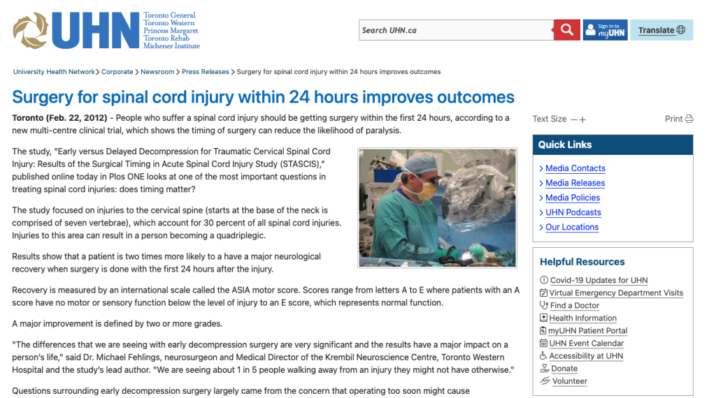
Surgery for spinal cord injury within 24 hours improves outcomes Toronto (Feb. 22, 2012) – People who suffer a spinal cord injury should be getting surgery within the first 24 hours, according to a new multi-centre clinical trial, which shows the timing of surgery can reduce the likelihood of paralysis. The study, “Early versus Delayed Decompression for Traumatic Cervical Spinal Cord Injury: Results of the Surgical Timing in Acute Spinal Cord Injury Study (STASCIS),” published online today in Plos ONE looks at one of the most important questions in treating spinal cord injuries: does timing matter? The study focused on injuries to the cervical spine (starts at the base of the neck is comprised of seven vertebrae), which account for 30 percent of all spinal cord injuries. Injuries to this area can result in a person becoming a quadriplegic. Results show that a patient is two times more likely to a have a major neurological recovery when surgery is done with the first 24 hours after the injury. Recovery is measured by an international scale called the ASIA motor score. Scores range from letters A to E where patients with an A score have no motor or sensory function below the level of injury to an E score, which represents normal function. A major improvement is defined by two or more grades. “The differences that we are seeing with early decompression surgery are very significant and the results have a major impact on a person’s life,” said Dr. Michael Fehlings, neurosurgeon and Medical Director of the Krembil Neuroscience Centre, Toronto Western Hospital and the study’s lead author. “We are seeing about 1 in 5 people walking away from an injury they might not have otherwise.” Questions surrounding early decompression surgery largely came from the concern that operating too soon might cause complications later on for the patient. But better surgical techniques and improved imaging capabilities over the last 20 years means that early surgery results in fewer complications. With consensus on early decompression growing among neurosurgeons, this study provides the scientific basis need to make policy changes that incorporates this evidence into a new standard of care for patients. “Since timing is such an important factor for treating spinal cord injuries we need to ensure that patients can get timely access to neurosurgical care,” said Dr. Fehlings. “This could mean the creation of neurosurgical centres of excellence, similar to stroke centres in Ontario.” There are 1,500 – 1,700 new injuries a year in Canada. Over a million people have an SCI in US in Canada and numbers are rising. According to the Rick Hansen Foundation, spinal cord injuries cost the system three billion dollars annually in Canada. Dr. Michael Fehlings is also a professor of neurosurgery at the University of Toronto. He holds the Krembil Chair in Neural Repair and Regeneration and is a McLaughlin Scholar in Molecular Medicine. About Krembil Neuroscience Centre The Krembil Neuroscience Centre (KNC), located at Toronto Western Hospital, is home to one of the largest combined clinical and research neurological facilities in North America. Since opening in 2001, KNC has been recognized as a world leader through its research achievements, education and exemplary patient care. The centre focuses on the advancement, detection and treatment of neurological diseases and specializes in movement disorders, dementias, stroke, spinal cord injury, blinding eye diseases, epilepsy and cancer-related conditions. For more information please visit www.krembil.com. About University Health Network University Health Network consists of Toronto General Hospital, Toronto Western Hospital, Princess Margaret Hospital and Toronto Rehabilitation Institute. The scope of research and complexity of cases at University Health Network has made it a national and international source for discovery, education and patient care. It has the largest hospital-based research program in Canada, with major research in cardiology, transplantation, neurosciences, oncology, surgical innovation, infectious diseases, genomic medicine and rehab. University Health Network is affiliated with the University of Toronto. Media Contact Phone: 416 340 4636Email: UHN.News@uhn.ca
U.S. and Canadian Scientists Form a Global Alliance for Nano-Bio-Electronics in Order to Rapidly Find Solutions for Neurological Disorders Such as Traumatic Brain Injury
LOS ANGELES, Feb. 13, 2012 /PRNewswire/ — The Society for Brain Mapping and Therapeutics (SBMT) announced today that the organization will hold its 9th Annual World Congress on Brain, Spinal Cord Mapping, and Image Guided Therapy from June 2-4, 2012 in Toronto, Canada. The world’s top brain and spinal cord scientists and surgeons will converge on the Toronto Metro Convention Center to find solutions to some of the most difficult to treat neurological disorders, including traumatic brain and spinal cord injuries, Parkinson’s Disease, Alzheimer’s Disease, and neurological cancers. The 2012 World Congress of SBMT is jointly supported by the American Association of Neurological Surgeons, the Government of Canada, the University of Toronto, and MaRS innovation; it is endorsed by the International Society for Magnetic Resonance Imaging in Medicine. The theme of this year’s World Congress is “Nano-Bio-Electronics,” which focuses on the integration of nanotechnology, stem cell research, and biomedical engineering, and imaging of the brain and spinal cord to make progress in the fight against neurological diseases. The aim of the Congress is to provide a multidisciplinary forum for health professionals in the fields of neurosurgery, neurology, psychiatry, radiology, neuroscience, engineering, as well as policymakers, to collaborate as a global alliance to rapidly advance treatment of neurological disorders. “The meeting will help us kick start a unique and efficient consortium, which will unite scientists and consolidate resources in order to help us quickly come up with solutions for the devastating neurological diseases affecting millions and costing billions in the US alone,” said Babak Kateb, Chairman of the Board of SBMT, President of the Brain Mapping Foundation, and Director of the National Center for Nano-Bio-Electronics (NCNBE). Dr. Kateb states, “The purpose of the Nano-Bio-Electronic alliance is to facilitate integration of nanotechnology, Stem cell and cellular therapy with medical devices and imaging. This consortium will impact global biomedical science and healthcare delivery through national and international partnerships with governments, universities, leading organizations and industries.” Among the notable participants of the 2012 World Congress includes Canadian Surgeon General Hans W. Jung, U.S. Navy Surgeon General Matthew Nathan, and Canadian Parliament Member Kirsty Duncan. Dr. Duncan, an advocate for brain research in Canada and a global voice for neuroscience initiatives, stated “I am honored to participate in this important conference. It is vital that we work to enhance our understanding of brain health through research and collaboration.” She added, “We must also affirm our commitment to improving the quality of life of those who live with a brain condition and of their families and informal caregivers.” Toronto was chosen for this year’s meeting because of the city’s strong and globally-connected network of neuroscientists, biomedical engineers, and investors in the biomedical and nanotechnology fields. Michael Fehlings, chairman of the local organizing committee, Professor of Neurosurgery, and Director of the Neuroscience Program at the University of Toronto, said “The meeting will showcase Canadian and international neuroscience talent in a broad range of disciplines and will highlight the latest advances in imaging, molecular and cellular mechanisms, bioengineering and surgical intervention.” Parimal Nathwani, Vice President of MaRS Innovation, added, “Forums like this represent an excellent opportunity for reviewing technologies and supporting collaboration across different institutions for more effective translation and commercialization opportunity.” The 9th Annual World Congress is still accepting abstract proposals for the meeting’s workshops, lectures, and presentation sessions. Abstract submission is open now until March 15th 2012. For the full list of 2012 speakers to register, or support of the 9th Annual World Congress of SBMT on Brain, Spinal Cord Mapping, and Image-Guided Therapy, please visit www.worldbrainmapping.org or call (310) 500-6196. Society of Brain Mapping and TherapeuticsSBMT is a non-profit society organized for the purpose of encouraging basic and clinical scientists who are interested in areas of Brain Mapping and Intra-operative Surgical planning to improve the diagnosis, treatment and rehabilitation of patients afflicted with neurological disorders. This society promotes the public welfare and improves patient care through the translation of new technologies into life saving diagnostic and therapeutic procedures. The society is committed to excellence in education, and scientific discovery. The society achieves its mission through multi-disciplinary collaborations with government agencies, patient advocacy groups, educational institutes and private sector (industry) as well as philanthropic organization. www.IBMISPS.org University of Toronto Neuroscience ProgramThe University Of Toronto Faculty Of Medicine established the U of T Neuroscience Program (UTNP) as a new academic program and appointed Professor Michael G. Fehlings as its first Director on September 1, 2008. The UTNP is a robust, integrated and collaborative academic program in neurosciences that leverages the unparalleled health science network at the University of Toronto, which includes U of T’s many departments and institutes, health science faculties, 9 fully-affiliated research hospitals and 20 community-affiliated hospitals and clinical care sites. MaRS InnovationMaRS Innovation provides an integrated commercialization platform that harnesses the economic potential of the exception discovery pipeline of 16 leading academic institutions in Ontario. MaRS Innovation is a not-for-profit organization with an independent industry- led board of directors, funded through the Government of Canada’s Networks of Centres of Excellence, the Province of Ontario through the Ministry of Research and Innovation, and contributions of its member institutions. Designed to enhance the commercial output of Toronto’s outstanding scientific research cluster, MaRS Innovation will make a significant contribution to Canada’s economic outlook and the quality of life for Canadians and others around the world. MaRS Innovation will advance commercialization through industry partnerships, licensing and company creation. The MaRS Innovation mission is to put Canada on the global innovation stage, by better connection of research with industry and strengthening Canada’s competitive capacity in the knowledge based business – in short, to launch a new generation of robust high growth Canadian companies. www.marsinnovation.com American Association of Neurological SurgeonsThe American Association of Neurological Surgeons (AANS) is the organization that speaks for all of neurosurgery. The AANS is dedicated to advancing the specialty of neurological surgery in order to promote the highest quality of patient care. http://aans.org SOURCE Society for Brain Mapping and Therapeutics
Timing for clinical trials for stem cell therapy in spinal cord injuries is right
OCTOBER 18, 2011 Timing for clinical trials for stem cell therapy in spinal cord injuries is right by Springer Regenerative medicine in spinal cord injuries (SCI) is proving to help the human body create new cell and nerve connections that are severed during this type of injury. In a review of current scientific research for stem cell treatment in SCI published this month in the Springer journal Neurotheraputics, Dr. Michael Fehlings and Dr. Reaz Vawda from the Krembil Neuroscience Centre, Toronto Western Hospital in Ontario, Canada, provide evidence that supports researchers moving beyond the lab to conduct human clinical trials for stem cells. Spinal cord injuries remain one of the most difficult conditions to overcome. Despite the advances made in surgical interventions, drugs and rehabilitation programs, the cascade of damage that ultimately affects the body at the cellular level cannot be reversed. Stem cell research brings tremendous hope to those who remain paralyzed after such a devastating injury. But according to Dr. Fehlings, patients are not able to realize the potential benefits of stem cell therapy because research is largely stuck in the laboratory. “With the exception of a few clinical trials, current research is stalled at the animal model stage,” said Dr. Fehlings. “Scientists from around the world have demonstrated as much as they can in lab models that stem cells have an impact on spinal cord injuries and can be transplanted into patients. Now we need the support and coordination of regulatory bodies to move this science forward.” The study critically evaluates 11 different cell types/sources and the evidence justifying their use. For example, BMSCs (Bone marrow stromal cells ) have an established safety record, as well as a beneficial effect after thoracic SCI. Glial restricted progenitors (GRPs) and oligodendrocytic progenitors (OPCs) are described to have a track record that favours the continuation of more clinical trials and Neural Progenitor Cells (NPC), of which one line of immortalized foetal NPCs has been subjected to extensive pre-clinical safety testing, may have potential benefits. Spinal cord injuries not only cause life-long disability and carry major psychological effects, injuries to the cervical spine (neck) have a mortality rate of 10 percent in the first year following injury and an expected lifespan of only 10 – 15 years post injury. Some of the evidence for increasing and expanding clinical trials from the study includes: “At this time, a strong patient advocacy base would likely help provide momentum to help translate current research into clinical applications,” said Dr. Fehlings. “Moving forward, all clinical trails must involve peer-reviewed assessment, regulation, independent monitoring, duplication, transparency and accurate record keeping of the every step of the process.” Dr. Fehlings acknowledges that no clinical intervention is 100 percent risk free and uses the examples of other novel therapies such as bone marrow transplantation and the polio vaccine to illustrate how science has forged ahead successfully, despite setbacks along the way. More information: Fehlings M, Vawda R (2011) Cellular Treatments for Spinal Cord Injury: The Time is Right for Clinical Trials. Neurotherapeutics. DOI 10.1007/s13311-011-0076-7 Provided by Springer