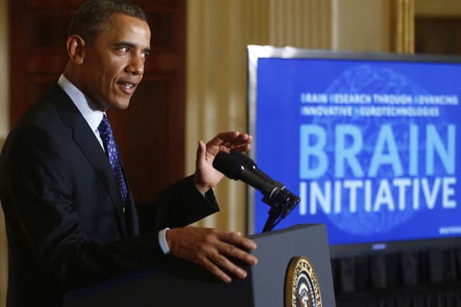
X2 Biosystems Presents at 5th Annual Brain Mapping Day at the US Congress
REDWOOD CITY, Calif. and WASHINGTON, April 21, 2016 /PRNewswire/ — X2 Biosystems, the leader in head impact safety solutions, today announced their participation at the 5th Annual Brain Mapping Day at the US Congress on April 20 in Washington, D.C. The event was organized by the Society for Brain Mapping and Therapeutics (SBMT), the Brain Mapping Foundation (BMF), the National Center for NanoBioElectronics, and the Congressional Neuroscience Caucus. During his presentation, X2 Chairman and CEO John Ralston, Ph.D., highlighted opportunities to correlate data from wearable head-impact sensors with computerized impact simulations and high resolution brain mapping to monitor, assess, and reduce the risks of brain injury in athletic and military environments. “We are honored to have this opportunity to support the efforts of SBMT and share our work with the Congressional Neuroscience Caucus,” said Dr. Ralston. “Combining X2’s wearable impact sensor solutions with high-performance data analytics platforms will enable significant breakthroughs in correlating real-time head impact data with multiple additional data streams, both at the network edge and in the cloud, to significantly enhance brain health and safety during athletic and military training exercises and sporting events.” The wide range of impairments to cognitive, motor, and sensory functions that can result from repetitive head impacts, often coupled with additional emotional and behavioral symptoms, has become recognized as an enormous public health challenge. Although much recent focus has been directed specifically at the issue of concussion injuries, a growing body of research is revealing that significant long-term brain injuries may result from cumulative head impacts even in the absence of clinically diagnosed concussion symptoms. In order to address this challenge, X2 Biosystems is developing solutions that combine the company’s “X-Patch” wearable head impact sensors with high-performance analytics platforms to address important goals such as: “We are grateful for the participation of industry leaders such as X2 Biosystems at the 5th Annual Brain Mapping Day,” said Aaron Filler, M.D., Ph.D., J.D., President of SBMT. “This is a fine example of government, industry, academia, and non-profit partnership to drive collaboration across multiple scientific and engineering disciplines, and to facilitate rapid introduction of life saving technologies for brain health, fitness, and therapeutics.” About X2 Biosystems (http://www.x2bio.com)X2 Biosystems merges wearable impact monitoring devices, neurocognitive assessment tools, wireless communications, and cloud data analytics to deliver the complete continuum of care in concussion management that athletic, medical, military, and industrial markets are now demanding in order to reduce the incidence, risks, and costs of head impacts and concussion injuries. X2 has offices in Redwood City, CA, and Seattle, WA. About The Society for Brain Mapping and Therapeutics (SBMT)(https://www.worldbrainmapping.org/about-sbmt) The Society for Brain Mapping and Therapeutics (SBMT) is a non-profit society organized for the purpose of encouraging basic and clinical scientists who are interested in areas of brain mapping, engineering, stem cell, nanotechnology, imaging and medical device to improve the diagnosis, treatment and rehabilitation of patients afflicted with neurological disorders. The society promotes the public welfare and improves patient care through the translation of new technologies/therapies into lifesaving diagnostic and therapeutic procedures. The society is committed to excellence in education and scientific discovery. The society achieves its mission through multi-disciplinary collaborations with government agencies, patient advocacy groups, educational institutes, industry, and philanthropic organizations. SOURCE X2 Biosystems Related Links http://www.x2biosystems.com
X2 Biosystems Awarded Pioneer in Healthcare Technology Award by The Society for Brain Mapping and Therapeutics
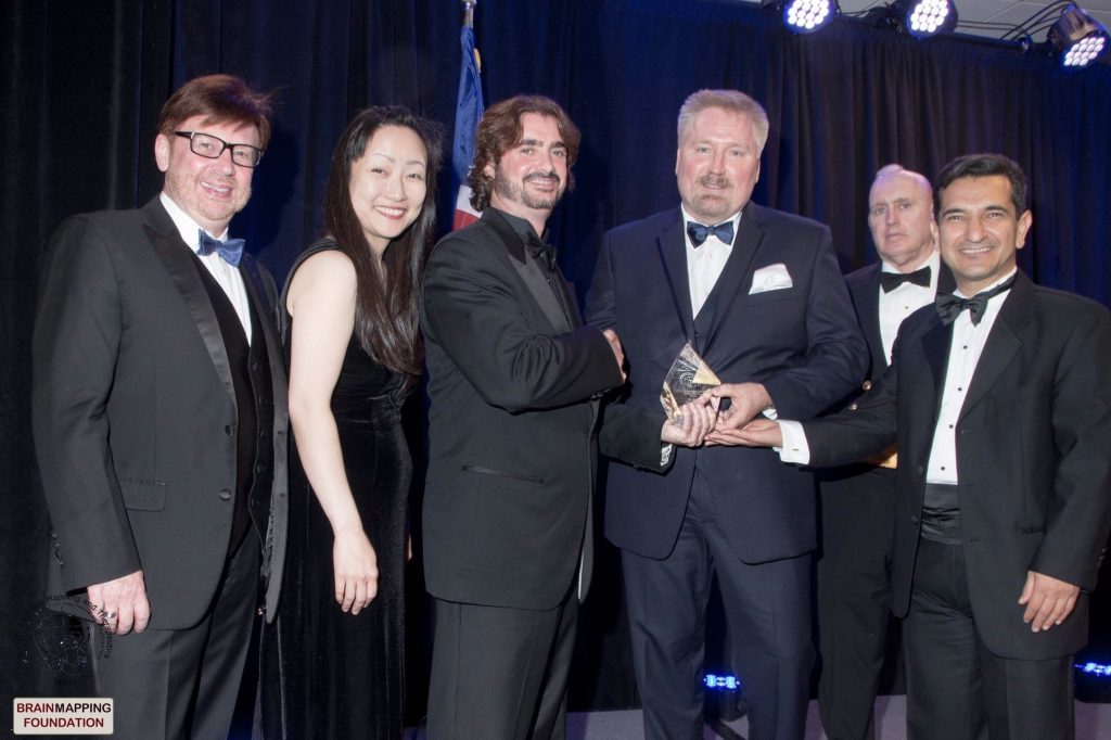
REDWOOD CITY, Calif. and MIAMI, April 18, 2016 /PRNewswire/ — X2 Biosystems, the leader in wearable impact monitoring devices and neurocognitive assessment tools, today announced that the company is the recipient of the The Society for Brain Mapping and Therapeutics (SBMT) 2016 Pioneer in Healthcare Technology Innovations Award for developing its next-generation head impact measurement sensor technology. The award was presented at a Saturday night gala at the 13th Annual SBMT World Congress in Miami, which featured Dr. Janet Kavandi Ph.D., astronaut and Director of the NASA Glenn Research Center, as keynote speaker. “We are deeply honored by this recognition from SBMT, an organization that brings together leading brain researchers across multiple disciplines from around the world,” said John Ralston, Ph.D. X2 Biosystems CEO. “This award highlights the significance of our technology and our collaborative efforts to reduce the risks, incidence and costs of head impact injuries.” “The SBMT Award Committee is truly pleased to select this year’s remarkable award recipients. We are honored to recognize their transformative discoveries and achievements in medical science, technology, and policy making,” said Vicky Yamamoto, Ph.D., a cancer scientist at Keck School of Medicine at USC, Chair of the Awards Committee, and Co-Chair of the Science Committee for SBMT. “X2 Biosystems’ solution stood out to the committee because of its truly innovative approach to head impact sensor technology. Along with previous award winners that include Siemens and Intel, X2 joins a very short list of technology innovators who have truly earned this world-class prestigious recognition from SBMT.” “SBMT is in the process of promoting global cross-disciplinary partnerships involving science, government and industry through its G20 World Brain Mapping and Therapeutics Initiative. This opportunity to honor X2 Biosystems is an exemplary validation of what SBMT hopes to accomplish through this global partnership with leaders of industry, academia, nonprofits and policymakers,” said Aaron Filler, M.D., Ph.D., J.D., President of SBMT and co-inventor of diffusion tensor imaging. X2 Biosystems was also given the honor of presenting on behalf of SBMT the 2016 Pioneer in Medicine Award to Dr. Bennet Omalu, who discovered the correlation between Chronic Traumatic Encephalopathy and the repetitive head trauma that is common to both athletes and soldiers. Although much recent focus has been directed specifically at the issue of concussion injuries, a growing volume of evidence is revealing that significant brain injuries may result even in the absence of clinically diagnosed concussion symptoms. X2’s “X-Patch” wearable impact sensors have become the world’s most widely deployed and tested head impact monitoring device, used in a continually expanding range of athletic activities from football (youth, high school, collegiate, pro) to hockey, soccer, lacrosse, rugby, Australian Rules Football, baseball, field hockey, wrestling, boxing, Taekwondo, mixed martial arts, skiing and BMX cycling. The X-Patch is also being actively evaluated for use in military training applications. Several landmark studies published by leading concussion researchers have demonstrated that safer training and playing techniques that incorporated the X-Patch achieved 30% to 70% reductions in the incidence of head impacts that can lead to concussion injuries.1-4 At the SBMT Annual Congress, X2 also presented initial results demonstrating that localized damage patterns observed in the brains of college athletes using high-resolution in vivo Diffusion Spectrum MRI (DSI), even in the absence of any diagnosed concussion symptoms, correlate with the impact loading recorded using X-Patch sensors. Multiple researchers are beginning to explore this application of the X2 sensors to help develop remove-from-play thresholds for both single head impacts and multiple head impacts accumulated over extended periods of time. “The brain is one of the final frontiers of medicine, with many mysteries still to be unlocked, and with X2 and its sensors, I believe that researchers now have a powerful set of tools to aid them in their quest to unravel the secrets of the brain, and medics have a tool to prevent brain trauma from accumulating,” said Dr. Harry Kloor, Ph.D. (physics), Ph.D. (chemistry), member of the SBMT Award and Science Committees. Click here to read the complete story about all of the awards presented at the 13th Annual SBMT Congress. X2 Biosystems CEO Dr. John Ralston will be speaking at the Congressional Briefing on Wednesday, April 20th at the 5th Annual Brain Mapping Day. The event will be held at the Rayburn House office building and was made possible by the Brain Mapping Foundation, Society for Brain Mapping and Therapeutics, National Center for NaoBioElectronics, SAP and X2 Biosystems. 1 UNH Helmetless Tackling Study2 Ole Miss Biosensing Study3 Journal of Neurosurgery Study 4 USA Football Heads Up Coaching Study ———– About X2 Biosystems (http://www.x2bio.com) X2 Biosystems merges wearable impact monitoring devices, neurocognitive assessment tools, wireless communications, and cloud data analytics to deliver the complete continuum of care in concussion management that athletic, medical, military, and industrial markets are now demanding in order to reduce the incidence, risks, and costs of head impacts and concussion injuries. X2 has offices in Redwood City, CA, and Seattle, WA. Media Contact: http://www.x2biosystems.com Contact: Michael RosenbergVP Marketing, X2 Biosystems(831) 818-2758michaelr@x2bio.com About The Society for Brain Mapping and Therapeutics (SBMT) (https://www.worldbrainmapping.org/about-sbmt) The Society for Brain Mapping and Therapeutics (SBMT) is a non-profit society organized for the purpose of encouraging basic and clinical scientists who are interested in areas of Brain Mapping, engineering, stem cell, nanotechnology, imaging and medical device to improve the diagnosis, treatment and rehabilitation of patients afflicted with neurological disorders. The society promotes the public welfare and improves patient care through the translation of new technologies/therapies into life saving diagnostic and therapeutic procedures. The society is committed to excellence in education, and scientific discovery. The society achieves its mission through multi-disciplinary collaborations with government agencies, patient advocacy groups, educational institutes and industry as well as philanthropic organization. Media Contact: Dr. Ken Green| Executive Director| Ken.Green@WorldBrainMapping.org Cell (202) 577-5105 Photo – http://photos.prnewswire.com/prnh/20160417/356240 SOURCE X2 Biosystems Related Links http://www.x2biosystems.com
Dr. Bennet Omalu, Congressman Andy Harris and Corporal Carpenter Receive Prestigious Recognition by a Leading BRAIN MAPPING ASSOCIATION, which has Launched and Supported GLOBAL BRAIN MAPPING INITIATIVES
NEWS PROVIDED BYSociety for Brain Mapping and Therapeutics (SBMT) 07 Apr, 2016, 07:35 ET MIAMI, April 7, 2016 /PRNewswire-USNewswire/ — The Society for Brain Mapping and Therapeutics (SBMT), the leading multi-specialty association for Brain Mapping, is holding its 13th Annual World Congress for Brain Mapping and Therapeutics in Miami, FL 8-10 April 2016 in partnership with Florida International University (FIU), the Brain Mapping Foundation, Children’s Hospital Miami, Uskudar University Turkey, the Institute for Nerve Medicine, and the California Neurosurgical Institute. Other strategic partners include National Aeronautics and Space Administration (NASA), the US Army’s Office of the Surgeon General, the Defense Advanced Research Project Agency (DARPA), the Department of State, Department of Energy, and the Los Alamos National Laboratory, the Department of Defense (DoD) and industry leaders: X2 BioSystems Inc., Surgical Theater, Medtronic, Stryker, EIZO , Haag-Streit USA, Time Medical, Nordic NeuroLab and Compumatics. The SBMT has been on the forefront of the global advocacy for BRAIN initiatives. Last year SBMT advocated and formulated African BRAIN Initiative, Middle East, and Malaysian BRAIN Initiatives to join brain initiatives already established in the US, Australia and Turkey, and also started a new SBMT chapter in Israel. This past year, SBMT leaders joined with Turkish scientists and physicians and held its 2nd Annual G20 Brain Mapping Initiative Summit in Istanbul and Ankara in Turkey jump starting Middle East-Turkey BRAIN Initiative. SBMT has been the global innovative leader in Brain Mapping, and has provided the model and impetus for the newly introduced International brain mapping course at the American Association of Neurological Surgeons (AANS), and a newly established initiative in brain mapping by the Institute for Electrical and Electronics Engineers (IEEE) organizations. The organization also has a special issue of Neurophotonics and Brain Mapping with SPIE-The International Society for Optical Engineering. “SBMT has been pushing for more global initiatives and global partnerships in last decade, and I am pleased to see that our advocacy has been fruitful,” said Aaron Filler, M.D., Ph.D., J.D. (13th President of SBMT) At the Annual Congress Gala Awards several individuals will be honored for their work and dedicated support of various brain research programs. Corporal William Kyle Carpenter, USMC (ret), recent and youngest living recipient of the Congressional Medal of Honor, is receiving SBMT’s Beacon of Courage and Dedication Award. The award committee is truly pleased to select these remarkable recipients this year. We are honored to recognize their transformative discovery and achievements in science, medicine, and policy making,” said Vicky Yamamoto, Ph.D., a cancer scientist at Keck School of Medicine of USC, Chair of the Awards Committee, and Co-Chair of the Science Committee. Ken Green, D.M.D. and Vicky Yamamoto, Ph.D. will be the recipients of the Golden Axon Leadership award. “Dr. Yamamoto has been devoting her last 13 years to SBMT’s global strategic and programmatic development; her leadership has been essential in advancing science and policy of SBMT,” said Babak Kateb, MD, Founding Chairman of the Board of SBMT. “Dr. Ken Green has been truly a great force behind our wounded warrior BRAIN Initiative and has been devoted to the SBMT cause for many years; we are grateful for their visionary leadership” he continued. Ruogu Fang, Ph.D. is the recipient of the Robin Sidhu Memorial Young Scientist Award. A Young Investigator Award will be selected and announced during the Gala Awards on April, 09/16. The National Space Biomedical Research Institute has also partnered with SBMT for its NSBRI-SBMT Young Investigator Award; the finalist will be announced at the meeting. The SBMT also has partnered with Haag-Streit USA and introduced a new clinical fellowship in skull base. Salman Abbasi Fard, M.D. is the recipient of the 2016 HS-SBMT Fellowship Award. “The Robin Sidhu Memorial Young Scientist Award is aimed at advancing the translation of information technology and supercomputing into neuroscience and brain discovery,” said Kuldip Sidhu Ph.D., member of the board of SBMT and Vice Chairman of the G20 World Brain Mapping Initiative The Scientific Program includes more than 200 presenters covering brain cancer, psychiatric disorders, neurodegenerative disorders, neurotrauma, neural repair, and regeneration, nanoneuroscience, cellular therapeutics, and immunotherapy, spinal disorders, engineering, brain policy and initiatives and radiation oncology. “The SBMT science committee invested 16 months in the preparation of this program, which is merely a snapshot of the scientific breadth of activities and achievements by our members,” said Dr. Ken Green, Executive Director of SBMT and co-chair of the awards and agenda committees. The membership, convention registration and gala registration are now open. Press Contact:Dr. Ken GreenExecutive Director of SBMTKen.Green@WorldBrainMapping.OrgCell: 202-577-5105 About SBMT:www.WorldBrainMapping.OrgAbout Brain Mapping Foundation:www.BrainMappingFoundation.Org SOURCE Society for Brain Mapping and Therapeutics (SBMT) Related Links http://www.WorldBrainMapping.Org
Society for Brain Mapping and Therapeutics Release: Leading Multispecialty Biomedical Association Presents Congresswoman Maxine Waters With Its Prestigious 2015 Pioneer In Healthcare Policy Award
Society for Brain Mapping and Therapeutics Release: Leading Multispecialty Biomedical Association Presents Congresswoman Maxine Waters With Its Prestigious 2015 Pioneer In Healthcare Policy Award Published: May 20, 2015 WASHINGTON, May 20, 2015 /PRNewswire-USNewswire/ — 12th Annual World Congress of Society for Brain Mapping and Therapeutics was held at the Los Angeles Convention Center in March but the impact of this world class scientific program has lasting effect on the policy, prevention, diagnosis and the treatment of neurological disorders such as Alzheimer’s disease, Brain injury, Brain Cancer and psychiatric disorders. Every year SBMT and Brain Mapping Foundation recognize policymakers who have significantly impacted prevention, diagnosis and the treatment of the Neurological disorder through pioneering legislations. Senators Ted Kennedy, Harry Reid and Barbara Boxer, Congresswomen Gabrielle Giffords and McMorris Rodgers, Congressmen Blumenauer, Fattah and Moran are among the past recipients of this prestigious award. In 2003 Congresswoman Waters introduced her first legislation on Alzheimer’s disease, which was aimed at creating a grant program to expand training and support services for families and caregivers of Alzheimer’s patients. Congresswoman Waters is also a champion of the Missing Alzheimer’s Disease Patient Alert Program, which is a small, cost-effective program aimed at helping local law enforcement officials quickly identify persons with Alzheimer’s disease who wander away from their homes and safely reunite them with their families. In December of 2014, Congresswoman Waters in partnership with Congressional Task Force on Alzheimer’s disease including Co-Chair Christopher Smith, advocated for a budget increase for Alzheimer’s Research. Her effort led to a $51 million increase in funding for Alzheimer’s research by President Obama in his budget proposal. This year, Congresswoman Waters has circulated a letter among her colleagues urging House appropriators to increase funding for Alzheimer’s research by $200 million in fiscal year 2016. She has also introduced a bipartisan resolution to make Alzheimer’s Disease an urgent National Priority. “Today we are recognizing one of the most influential members of the Unite States Congress who has impacted the care of millions of patients with Alzheimer’s disease through her pioneering legislation,” said Dr. Babak Kateb, Chairman of the Board of Society for Brain Mapping and Therapeutics, President of Brain Mapping Foundation, Director of National Center for NanoBioElectronics, Neuroscientist, Department of Neurosurgery, Cedars Sinai Medical Center. The Society for Brain Mapping and Therapeutics has been on the forefront of scientific research and translation of advance technologies into clinical neuroscience by engaging scientists and engineers from NASA, Los Alamos National Labs and consortium of neurosurgeons, neurologists, radiologists, nanotechnologists, stem cell scientists and engineers. The Society has been a great supporter of the Congresswoman’s vision and efforts. “I want to thank the Society for Brain Mapping and Therapeutics for honoring me with this award,” said Congresswoman Waters. “I also appreciate the Society’s commitment to improving our understanding of Alzheimer’s disease and other diseases affecting the brain. The Society’s dedication to finding cures for these diseases will help us immensely in our collective efforts to alleviate human suffering.” The award was presented to the Congresswoman at the 4th Annual Brain Mapping Day at the US Congress, Congress Visitor center, HVC 201 on May 19th 2015. The following was the scientific schedule of the 4th Annual Brain Mapping Day where the award was presented: 10:20am-10:30am: Overview of Los Alamos National Lab Technology and potential application in Brain Mapping and Therapeutics, Nancy Sauer, Ph.D., Associate Director for Chemistry, Life, and Earth Sciences, Los Alamos National Laboratory 10:30am-10:40am: Ultrasonic Technology from Bomb to Brain Discovery, Dipen N. Sinha, PhD, Laboratory Fellow & Team Leader, Los Alamos National Laboratory 10:40am-10:50am: Neuroimaging at Los Alamos National Laboratory, Michelle Espy, PhD, Research Scientist & Team Leader, Applied Physics Lab, Los Alamos National Laboratory 10:50am-11:00am: SBMT-South Africa Brain Mapping Initiative, The Honorable Monwabisi Bevan Goqwana, Ph.D., Member of National Assembly (Parliament) of South Africa The program was moderated by Dr. Babak Kateb & Actor and Neuroscience Advocate, Robert Picardo and jointly sponsored by Congressional Neuroscience caucus, Brain Mapping Foundation, SBMT, National Center of NanoBioElectronics and Medtronic. Professors Benjamin Aribisala (Nigerian delegate) and Nevzat Tarhan and Mr. Serdar Karagoz (Turkish delegates) were amongst the SBMT members who presented the award to the honorable on behalf of the SBMT membership and board of directors. Next year’s Annual World Congress of SBMT is scheduled for April 7-9, 2016 in Miami. About SBMT:WWW.WorldBrainMapping.Org Media Contacts Bryan ArozBryan.Aroz@WorldBrainMapping.orgTel: 310-500-6196 Photo – http://photos.prnewswire.com/prnh/20150520/217391 To view the original version on PR Newswire, visit:http://www.prnewswire.com/news-releases/leading-multispecialty-biomedical-association-presents-congresswoman-maxine-waters-with-its-prestigious-2015-pioneer-in-healthcare-policy-award-300086379.html SOURCE Society for Brain Mapping and Therapeutics
From Outer Space to Innerspace: The Society for Brain Mapping and Therapeutics

Dr. Babak Kateb, Founding Chairman of the Board of the Society for Brain Mapping & Therapeutics and Director of National Center for NanoBioElectronics Nathaniel Gore of PLOS Collections, on the Neuromapping and Therapeutics Collection and the upcoming SBMT Annual Congress. The Society for Brain Mapping and Therapeutics (SBMT) is a multidisciplinary and multispecialty non-profit biomedical association organized for the purpose of encouraging basic and clinical scientists who are interested in areas of Brain Mapping, engineering, stem cell, nanotechnology, imaging and medical device to improve the diagnosis, treatment and rehabilitation of patients afflicted with neurological disorders. Founded in 2003 with the aim of filling the educational gap between biological and physical science disciplines, the organization came about after a successful collaboration between NASA/JPL, Keck School of Medicine of USC, and Caltech. Since then, the Society has held 13 annual world congresses in the US, France, Canada and Australia, established its own Brain Atlas, published the inaugural textbook of nanoneurosurgery and been a driving force behind President Obama’s BRAIN initiative. In 2014, the Society launched the G20 World Brain Mapping and Therapeutics conference in Brisbane in order to develop global cooperation and partnership for brain discovery and repair. The Society is a leader in the field of Brain Mapping and Therapeutics and could be considered as the world’s fastest growing translational neuroscience think-tank, with near 4,000 members – consisting of scientists, surgeons, physicians and engineers – focussing their attention on narrowing the gap between disciplines. The organization helps to bridge these gaps through annual Conventions and satellite symposia, the SBMT University, journals, prestigious awards, Brain Mapping Days at the US Congress, partnership with National Center for NanoBioElectronics (NCNBE) for translating, integrating and commercializing of advance technologies into clinical trials , student Chapters and its global partnerships with G20 and African Brain Mapping initiatives. It’s due to this shared cross-disciplinary nature that PLOS ONE and SBMT have, since 2010, partnered on the Neuromapping and Therapeutics Collection. The aim of the Collection is to provide a forum for interdisciplinary research aimed at translation of knowledge across a number of fields such as neurosurgery, neurology, psychiatry, radiology, neuroscience, neuroengineering, nanoneuroscience/nanoneurosurgery and healthcare policy issues that affect the treatment delivery and usage of certain devices, drugs and imaging technologies. PLOS ONE’s wide scope and broad publication criteria make it a perfect venue to publish and collate relevant articles in these areas of research into one cross-disciplinary collection. Our hope is that by encouraging and facilitating further research, replication, and sharing of both positive and negative results, this Collection will become a catalyst for continued innovation and discovery in brain mapping and therapeutics. Join us at the Upcoming 12th Annual SBMT Congress PLOS will be attending the 12th Annual Congress of SBMT in Los Angeles on March 6-8 and look forward to discussing the Neuromapping and Therapeutics Collection and the PLOS Neuro Community site. In the coming weeks, society members will be blogging about their research and why they value being a member of SBMT. Abstract submissions for the Congress are still open, as are nominations for the Young Investigator Award – the deadline is February 9th. Connect with SBMT through Facebook, Twitter or LinkedIn accounts. For more information about the collection – or PLOS Collections in general – contact collections@plos.org ————————————————————————— Dr. Babak Kateb (@BabakKateb) is the Founding Chairman of the Board of The Society for Brain Mapping & Therapeutics, Research Scientist, Department of Neurosurgery, Cedars Sinai Medical Center and Director of National Center for NanoBioElectronics Nathaniel Gore (@thengore) is the Editorial Project Manager of PLOS Collections
Society for Brain Mapping and Therapeutics holds its 12th Annual World Congress at the LA Convention Center in March announcing its African Brain Mapping Initiative
LOS ANGELES, Jan. 15, 2015 /PRNewswire-USNewswire/ — World leading scientists, engineers, physicists, physicians and surgeons converge to Los Angeles Convention Center from Friday March 6th to Sunday March 8th in order to rapidly identify, translate, integrate and commercialize advanced technologies, which could help brain discovery and assist with diagnosis and treatment of neurological disorders such as PTSD, brain trauma, brain cancer, neurodegenerative disorders, mental disorder, spinal disorders and peripheral nerve injuries. The 12th Annual World Brain Mapping and Therapeutics convention will have 550 speakers, 110+ scientific sessions and 10 keynote speakers including Congressman Chaka Fattah and Chief of the US Army General Ray Odierno. “Since its inception, the Society for Brain Mapping and Therapeutics has been a pioneer in research, advocacy, and service to the medical research community. Los Angeles is proud to host this gathering of researchers, physicians, policy makers, scientists, and industry leaders to advance scientific and technological discoveries of the human brain.” said Los Angeles Mayor Eric Garcetti. Last June, SBMT launched the G20 World Brain Mapping and Therapeutics Initiative in Italy and held its first annual G20 World Brain Mapping and Therapeutics Symposium in Brisbane, Australia to mobilize a global partnership for brain disease prevention, discovery and treatment. Brain Mapping Foundation, National Center for NanoBioElectronic, Amen Clinics, and Compumedics were amongst the supporters of the G20 event. “SBMT has been on the forefront of multidisciplinary brain science, technology,and discovery and in the last two decades through a global multidisciplinary partnership and collaboration” said, Dr. Pantaleo Romanelli, President Elect of SBMT, Chief Scientist of AB Medica, Consultant and Scientific Director, Brain Radiosurgery, Cyberknife Center, in Milan, Italy, Visiting Scientist, European Synchrotron Radiation Facility (ESRF), in Grenoble, France African Brain Mapping and Therapeutics Initiative was launched as part of the G20 Brain Mapping Initiative, which aimed at complementing President Obama’s BRAIN Initiative and EU Human Brain Project. In September 2014, Dr. Babak Kateb, Chairman of the Board of Directors of SBMT met with Drs. Glenda Gray, President and CEO of South Africa Medical Research Counsel, John Ouma, Chairman of the Department of Neurosurgery, University of the Witwatersrand, Marlon Burges, Chairman of Medical Device Association of South Africa and the local prominent scientists and leaders in South Africa discussing the details of the initiative. “We thank SBMT and Dr. Kateb for visiting us in South Africa and engaging us in this Initiative. We are very excited to be a strategic partner of SBMT and help this initiative by engaging the strategic groups in the entire Africa including the African Academy of Science,” said Dr. John Ouma, Member of the Board of SBMT, Chairman of the Department of Neurosurgery, University of the Witwatersrand, Johannesburg, South Africa and Secretary General of South African Neurosurgical Association. The topics of this year’s World Congress include: brain trauma, mental health, brain policy and research funding, engineering, brain cancer, nanoneurosurgery, metadata and brain mapping, medical imaging, device, material science, radiation oncology, deep brain connectomics and spinal disorders and diagnostics. “This year we particularly emphasized on global partnership with foundations and industry leaders; we have reached out to near 300 foundations and near 1200 industry leaders on our network with the aim of creating a united front on advancing brain discovery. “As the 12th President of SBMT, I made advanced technology and engineering a core for the scientific program and our global initiatives,” said Dr. Shouleh Nikzad, 12th President of SBMT, Senior Research Scientist and Lead of the Advanced UV/Vis/NIR Detector Arrays, Imaging Systems, and Nanoscience Group, at NASA’s Jet Propulsion Laboratory, California Institute of Technology, CA, USA. The 12th annual World Brain Mapping of SBMT is still accepting abstracts, and the organization is planning to expand its membership from 4000 to 6000 in next year and has introduced a multidisciplinary Young Investigator award in partnership with National Space Biomedical Research Institute (NSBRI) in order to encourage younger scientists to join the organization. For more information about how to become a member or register for the convention, please visit: www.WorldBrainMapping.org About SBMT: www.WorldBrainMapping.org Media Contact:Dr. Vicky YamamotoTel: 310-500-6196Vicky.Yamamoto@med.usc.edu Photo – http://photos.prnewswire.com/prnh/20150115/169420 SOURCE Society for Brain Mapping and Therapeutics Related Links https://www.worldbrainmapping.org
Brain Mapping Leader Says Conference Planning Can Change the World
The annual conference for the Society for Brain Mapping & Therapeutics (SBMT) began a dozen years ago as a small summit for 20 doctors, scientists and engineers who came together to discuss advances in neuroscience. Today, the conference brings together a global community of thousands of specialists in the fields of neuroscience, engineering, neurosurgery, psychiatry, psychology, molecular biology, neurology, radiology and oncology. One of the unique aspects of this conference is all of those different disciplines learning and working together, which is unusual in the scientific community. The interdisciplinary nature of the program is also why the Society is making such a profound impact on the study of nanoscience (science at the atomic and molecular level), with many important real world implications. The best way to explain brain mapping in laymen’s terms is by using Google Maps as an analogy. Traditional brain scanning is like Google Maps zoomed out until you can see the shape of countries. Today’s leading technology in brain mapping is like Google Maps Street View, where you can see cars on the street, which equates in this case with probing deep into the brain’s molecular level. Furthermore, it’s becoming a primary issue for world governments in advanced countries. Over the last couple of years, there’s been a global push among G20 nations to speed up the study of neuroscience, with governments dedicating millions of new dollars for research. President Obama’s BRAIN Initiative, launched in 2013, provides over $300 million earmarked through 2015. According to the White House website, the BRAIN (Brain Research through Advancing Innovative Neurotechnologies) Initiative is “a bold new research effort to revolutionize our understanding of the human mind and uncover new ways to treat, prevent, and cure brain disorders like Alzheimer’s, schizophrenia, autism, epilepsy, and traumatic brain injury.” Presently, the World Health Organization estimates that about a third of the worldwide adult population suffer from a mental disorder such as depression, anxiety and schizophrenia. If also taken together with neurological disorders, such as dementia and stroke, these “disorders of the brain” account for 13% of the global disease burden. There is also an exponential increase in psychological trauma among military veterans in the U.S. That is why the SBMT conference is gaining so much exposure. One of the primary outcomes from the conference to date is the publication of the first academic textbooks in the fields of nano-neurosurgery and nano-neuroscience. SBMT members have also published a Brain Anatomy Atlas and Brain Disorder Atlas to assist with brain navigation during surgery. “The textbooks are major breakthroughs…. Just think about the first textbooks for biology,” says Dr. Babak Kateb, founding chairman of the board of SBMT, and president of the Brain Mapping Foundation. “And in terms of policy, we have been a force behind President Obama’s BRAIN Initiative, and we have been successful helping wounded warriors through all of this specialty research.” In a further effort toward global cooperation, the SBMT and European-based Human Brain Project entered a joint partnership to develop the G20 World Brain Mapping and Therapeutics conference in Brisbane last month. “The idea is we bring together G20 nations and create a global cooperation on brain mapping and therapeutics, which is a direct result of our convention,” says Dr. Kateb. “So when you’re talking about the social legacy of a convention, or gold standard of a convention, this is a very young organization that started with just 20 people. When people are creating an event, it is very important for them to understand the potential of conference planning to set up a gold standard in their field.” Some of the new medical innovations that SBMT members have pioneered include new retinal imaging to diagnose Alzheimer’s disease, and new nano-drugs to treat brain diseases. SBMT scientists have even partnered with NASA to develop new microwave devices to treat different cancers, which have far less harmful side effects than traditional radiation therapies. In an attempt to build on the event’s social legacy, Dr. Kateb and the SBMT leadership are expanding the reach of the Society’s membership and collective knowledge base by developing Brain Mapping Initiatives in Europe, Asia/Pacific, Africa and the Middle East. The overall goal is to increase the direct impact on communities worldwide where SBMT members are based. Past SBMT annual conferences have taken place in Sydney, Toronto, Baltimore, San Francisco, France, Harvard University and Bethesda, among others. Dr. Kateb has also traveled to South Africa to meet with scientists and doctors in Cape Town, and to research the destination as a possible venue for a future conference. “When you bring thought leaders together—5, 10, 20 of them—if you’re persistent, you can create a shift in the entire way of thinking in any large organization,” says Dr. Kateb. “We have accomplished that in science, and science is pretty hard to change.”
G20 World Brain Mapping Initiative/Summit in Australia started with important messages of Collaboration from Congressman Chaka Fattah, Congressman Earl Blumenauer and Member of the Canadian Parliament Kirsty Duncan and pioneering work by the top G20 scientist
BRISBANE, Australia, Nov. 13, 2014 /PRNewswire/ — Earlier this year in Milan, Italy, the Society for Brain Mapping & Therapeutics (SBMT) announced its G20 World Brain Mapping and Therapeutics Initiative. Today, the Amen Clinics, Compumedics Inc, SBMT, and BMF hold the first annual summit on G20 World Brain Mapping and Therapeutics Initiative in Brisbane, Australia on Nov. 13th in Mercure Hotel. “We have spent more than 3 years to formulate this initiative, which we announced it earlier this year in Milan and held the 11th Annual World Congress in Sydney this year in order to finally bring the G20 World Brain Mapping summit” said Dr. Kuldip Sidhu, Professor of Stem Cell at UNSW, the Past President of SBMT and a current President of SBMT-Australia. The summit will start with messages of cooperation from the U.S. Congressman Fattah, the U.S. Congressman Blumenauer (Chairman of the Congressional Neuroscience Caucus) and Member of the Canadian Parliament Kirsty Duncan. These leaders encourage all scientists to work together to curb the growing cost associated with diagnosis and treatment of neurological disorders through introducing game-changing diagnostics and therapeutics. The summit program includes talks from top US, Australian, Italian, Turkish, and European Brain Mapping Initiative scientists covering topics such as advanced imaging in diagnosis of Alzheimer’s disease, psychiatric disorders, brain cancers, neurodegenerative disorders, big data in brain mapping, strategies for global clinical trials, policies that could facilitate translation, integration and commercialization of devices and therapeutics such as nanoneurosurgery/nanoneuroscience, neurotrauma, and military medicine, as well as a roundtable discussion with the US and Canadian Policymakers. “Specifically, the goals of the program are to: Build solid working collaboration between the USA, Australia and other G20 nations on translational, clinical neuroscience including all aspects of the Brain Mapping and Therapeutics; Build strategic industry-academia-government alliance for brain discovery, disease prevention, diagnosis, and treatment; Facilitate rapid integration, translation, and commercialization of innovations in the field of Brain Mapping & Therapeutics worldwide; Launch complementary Brain Mapping Initiatives across the G20 nations; Facilitate clinical trial and formulation of joint Institutional Review Board taskforce across G20” said, Dr. Babak Kateb, Chairman of the Board of the SBMT and Research Scientist, Department of Neurosurgery, Cedars-Sinai Medical Center. “I am very happy that Australia took the lead on this topic and already planning the 2016 G20 World Brain Mapping Summit in Turkey with our Turkish colleagues.” Media Contact: Bryan Aroz: Bryan.Aroz@WorldBrainMapping.org Tel: 310-9808340 About SBMT: WWW.WorldBrainMapping.org SOURCE Society for Brain Mapping and Therapeutics Related Links http://www.WorldBrainMapping.org
G20 World Brain Mapping and Therapeutics Initiative by SBMT will include significant partnership with European Human Brain Project.
MILAN, June 26, 2014 /PRNewswire-USNewswire/ — According to the World Health Organization’s large-scale studies, about a third of the adult worldwide population suffer from a mental disorder such as depression, anxiety and schizophrenia. If also taken together with neurological disorders, such as dementia and stroke, these “disorders of the brain” account for 13% of the global disease burden. This surpasses both cardiovascular diseases (5%) and cancer (10%). Countries with the highest rate of burden > 650 Disability Adjusted Life Years [DALYs] per 100,000 population included the USA, UK, Russia, and Australia. The annual cost of taking care of patients with neurological disorders in the US alone approaches $400B of which $200B is for Alzheimer patients. We believe that this cost in 10 years could reach to $1T in the US alone. In China 975,000 people die annually from brain trauma alone and the Alzheimer population in the Australasia corridor is rapidly on the rise. Thus, European Union, China, India, Japan and Australia are not immune from such high cost of healthcare despite having 100% government insurance. About 3 million Australians are estimated to experience symptoms of a mental disorder (ABS 2008). Australian Institute of Health and Welfare’s (AIHW) estimates that over $6 billion per annum is spent on mental health-related services in Australia. From the European Brain Council study, it is estimated that the annual cost of brain disease in Europe was 798 billion Euros in 2010. The total estimated worldwide costs of dementia were US$604 billion in 2010 and growing to be a near $1T annually. About 70 percent of the costs occur in Western Europe and North America. An US government study, the National Comorbidity Study Replication, estimated that serious mental illness accounted for $193 billion in lost earnings. The G20 World Brain Mapping and Therapeutics was announced initially at the 12th Annual World Congress of SBMT in Sydney, Australia earlier this year but the partnership with EU-Human Brain Project was announced yesterday at the 11th Annual meeting of “the future of Healthcare,” which was held at a one of the most historic venues in Milan/Italy, the Four Seasons, on June 25, 2014; thanks to the AB Medica’s generous sponsorship. “We are truly delighted to have top European and US scientists in this convention, which is designed to predict the future of healthcare based on advance science/technology and very happy that SBMT now will be closely partnering with EU Human Brain Project,” said Dr. Pantaleo Romanelli, newly elected President of European Chapter of Society for Brain Mapping & Therapeutics (SBMT), Chief Medical Officer, AB Medica, Milano, Italy, Consultant and Scientific Director, Radio-neurosurgery, CDI, Milano, Italy, Visiting Scientist, European Synchrotron Radiation Facility, Grenoble, France. “SBMT is a world leader in the field and has been the global voice for translational neuroscience. We continue to be on the forefront of pioneering medical research, advocacy as well as global partnership,” said Dr. Kuldip Sidhu, the Past President of SBMT, A/Prof. of Stem Cell at University of New South Wales and President of SBMT-Australia. The European Commission has officially announced the selection of the Human Brain Project as one of its two Flagship projects. The new project will federate European efforts to address one of the greatest challenges of modern science: understanding the human brain. The cost is estimated at 1.19 billion Euros. “Global collaboration is the key to advancing neuroscience to understand the human brain from genes to cognition. It is critical to integrate and coordinate both basic and clinical sciences to bring their insights to benefit the patient,” said Dr. Sean Hill, Co-Director of Informatics Research at EU Human Brain Mapping Project. President Obama recently added 100-million-dollars to the FY14 and $200M FY15 budget of the US government as part of the BRAIN initiative. This is an additional new funding for the NIH, DARPA, and NSF. Similarly a recent the Prime Minster of Australia, Toy Abbott, added $200 million dollar to his federal budget in order to combat dementia in addition to substantial more funding for medical research. “I am glad that our colleague in European Brain Project have understood the importance of collaborating with SBMT, which is a leading edge organization for Brain Mapping & Therapeutics,” said Dr. Babak Kateb, Founding Chairman of the Board of SBMT, President of Brain Mapping Foundation, and Research Scientist at the Department of Neurosurgery, Cedars-Sinai Medical Center in Beverly Hills, California, USA; “we believe there is a great need for a global action focused on: 1) more systematic and methodical study of brain in human health in a consortium approach; 2) well coordinated global response to the rising burden with neurological disorders and global harmonizing of the related policies; 3) well planned neuro-economical assessment of the future impact of disease, diagnostics and prevention; 4) facilitating translation of technologies across disciplines of science in order to rapidly identify and introduce new generation of therapeutics; 5) unifying global regulations and guidelines on clinical trials and drug/device-combination discovery; 6) establish global partnership and new funding initiatives across academic, educational, industry and non-profit organizations and 7) facilitate integration, translation and commercialization of neurotechnologies such as nanoneuroscience, cellular therapeutics, imaging and advance electronics/devices,” he continued. Australia is hosting the Group of Twenty (G20) developing nations in November of this year in Brisbane. This is a great opportunity for the heads of states of the G20 Nations to potentially put the G20 World Brain Mapping & Therapeutics initiative introduced by the SBMT and Brain Mapping Foundation on their current and future agenda. This year SBMT is partnering with Australian American Association, Research Australia, Federal and State governments of Australia in order to hold the first annual G20 World Brain Mapping and Therapeutic summit in Australia on Nov. 12th 2014. About SBMT: For more information please visit: www.WorldBrainMapping.org About AB Medica SPA: For more information about AB Medica SPA please visit: http://www.abmedica.it/ Media Contact: Bryan Aroz| Society for Brain Mapping & Therapeutics (SBMT)8159 Santa Monica Blvd. Suite #200|West Hollywood,CA90046|Tel: (310) 500-6196|Fax: (323) 654-3511| Bryan.Aroz@WorldBrainMapping.orgpress@WorldBrainMapping.org SOURCE Society for Brain Mapping and Therapeutics
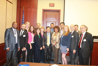
Dr. Ramin Rak Speaks at U.S. Congressional Briefing About Brain Mapping During Awake Brain Surgery
Dr. Ramin Rak Will Be One Of 10 Experts From Across Nation To Speak, As Part Of Congressional Briefing On Brain Mapping And Neuroscience Third Annual Brain Mapping Day at the US Congress, May 20th 2014 A Congressional Brief on the state-of-the-art in Brain Mapping, Therapeutics & Translational Multi-speciality Neuroscience, Organizers: Society for Brain Mapping and Therapeutics (SBMT), Brain Mapping Foundation and Congressional Neuroscience Caucus 8:10-8:15Award Presentation to the Honorable Congressman Fattah 8:20-8:352014 SBMT-BMF Pioneer in Healthcare Policy Award RecipientCongressman Chaka Fattah, The United States Congressman from 2nd District of Pennsylvania, USA 8:40-8:45New Ways to Measure Brain ActivityGeoff Ling, MD, PhD, FAAN, Director of Biological Technologies, Defense Advance Research Project Agency (DARPA), Professor and Acting Chair of the Department of Neurology, Uniformed Services University of the Health Sciences (USUHS), USA 8:45-8:50Mapping Brain Function – A Key to Finding Effective Treatments for TBI and PTSDTimothy J. O’Leary, MD, PhD Acting Chief R&D Officer The US Department of Veterans Affairs, Office of Research & development, USA 8:50-8:55Neuroimaging of Brain Injury in the US MilitaryJames P. Kelly, MA, MD, FAAN, FANA Director, National Intrepid Center of Excellence (NICoE) Walter Reed National Military Medical Center Clinical Professor of Neurosurgery University of Colorado School of Medicine, USA 8:55-9:00Ambulance Based Treatment for Acute StrokeEric M. Bailey, Ph.D. Founder & CEO, Neurologica Corporation, USA 9:00-9:05Creating Windows into the Brain: The Emerging Synergy of Neuroscience and Medical DevicesTimothy Denison, Ph.D. Director of Core Technology, Technical Fellow, Medtronic Corporation, USA 9:05-9:10Brain Mapping During Awake Brain Surgery Ramin Rak, M.D., F.A.N.S.Attending Neurosurgeon, North Shore-LIJ Health Systems, Winthrop University Hospital, Catholic Health System, Long Island, NY; SBMT Board Member; Director, Brain Tumor Program, North Shore-LIJ Huntington HospitalCo-Surgical Director& Director Of Awake Craniotomy & Brain Mapping Program, Long Island Brain Tumor Center, USA 9:10-9:15Los Alamos Contributions to Next Generation Brain Mapping and TherapeuticsDavid Pesiri, PhDDirector, The Richard P. Feynman Center for InnovationLos Alamos National Laboratory, USA 9:15-9:20Functional imaging in the pediatric brainMichael R. Yochelson, MD, MBAVice President of Medical Affairs & Chief Medical Officer MedStar National Rehabilitation Hospital, SBMT Board MemberVice Chair of Clinical Affairs, Department of Rehabilitation Medicine MedStar Georgetown University HospitalProfessor, Clinical Neurology & Clinical Rehabilitation Medicine Georgetown University, USA 9:20-9:25The Cyberbrain project: monitoring and modulation of brain function using wireless brain electrodesPantaleo Romanelli, MDScientific Director, AB Medica, SBMT Board Member Scientist, European Synchrotron Radiation Facility (ESRF), Grenoble, France Consultant Neurosurgeon and Scientific Director, CDI, Milano, Italy 9:25-9:30Impact of Neurological Disorders on the World Economy, A $1T Question?Kirsty Duncan, PhD, Member of Parliament of Canada Professor of Health Studies at University of Toronto Member of Board of Directors of SBMT, Recipient of 2012 SBMT-BMF Pioneer in Healthcare Policy Award Moderator:Dr. Babak Kateb | Founding Chairman of the Board of Directors|CEO and Scientific Director Society for Brain Mapping & Therapeutics (SBMT)| Director of National Centre for Nano-Bio-Electronics (NCNBE)| Scientist| Maxine Dunitz Neurosurgical Institute| Department of Neurosurgery| Cedars Sinai Medical Center| Chief Editor| SBMT-PLoSOne NeuroMapping & Therapeutics| Editor of The Textbook of Nanoneuroscience and Nanoneurosurgery, President and Scientific Director of Brain Mapping Foundation, 9:35-10:00 Discussion Program End Sharp at 10AM EST https://www.worldbrainmapping.org/2nd-annual-brain-mapping-day