Challenges in Interpreting DTI Data
Challenges in Interpreting DTI Data: Addressing Noise, Artifacts, and Limitations in Accuracy
Objective: To explore the challenges and limitations in interpreting DTI data, including addressing noise, artifacts, and accuracy concerns.
Challenges in DTI Data Interpretation:
- Noise and Artifacts:
- DTI data is prone to noise due to patient motion, magnetic field inhomogeneities, or scanner limitations. These factors can introduce errors in the reconstructed images and misinterpret the true white matter pathways.
- Common Artifacts:
- Motion Artifacts: Caused by patient movement during scanning.
- Partial Volume Effects: Occur when a voxel contains a mixture of tissue types, which can affect diffusion estimates.
- Susceptibility Artifacts: Caused by variations in magnetic susceptibility, often near air-tissue interfaces.
2. Low Spatial Resolution:
- While DTI provides excellent information about white matter pathways, the spatial resolution is limited. This can result in difficulty detecting smaller tracts or resolving complex brain regions.
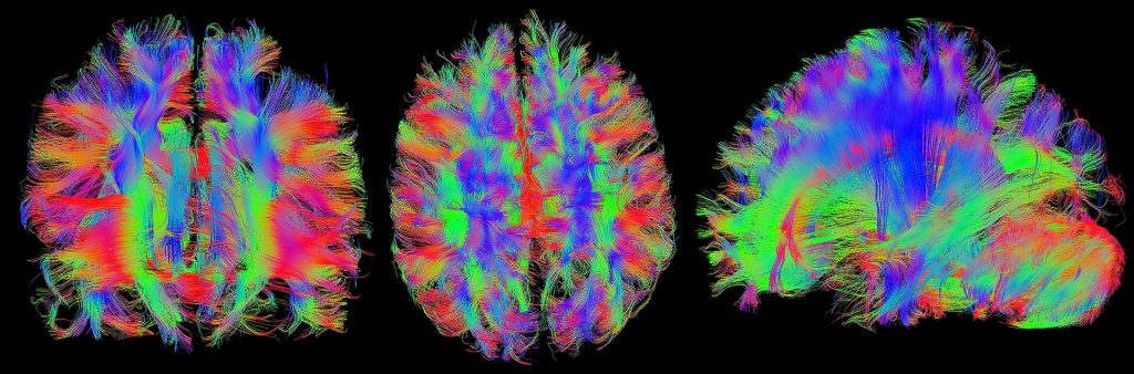
3. Complexities in Fiber Orientation:
- In areas where fibers are crossing or orienting in multiple directions, DTI can struggle to resolve these complex configurations accurately. This is a limitation when trying to track fibers in regions like the corona radiata or regions with fiber crossing.
Solutions to Improve Data Interpretation:
- Higher-Resolution Imaging:
- Using high-field MRI scanners or advanced acquisition techniques can help improve the resolution of DTI data, reducing the impact of partial volume effects.
2. Advanced Reconstruction Algorithms:
- New algorithms such as constrained spherical deconvolution (CSD) can resolve crossing fibers more accurately, improving tractography results.
3. Post-Processing Methods:
- To reduce noise and artifacts, various post-processing techniques like denoising, motion correction, and quality control are employed in DTI studies.
Real-World Example:
In patients with Multiple Sclerosis (MS), DTI has been used to detect changes in white matter integrity. However, due to the complexities in fiber orientation and the presence of lesions, interpreting DTI data in MS patients requires careful consideration of these challenges. Combining DTI with other imaging techniques, such as magnetization transfer imaging (MTI), can provide a more comprehensive assessment of white matter integrity in MS.
Case Study:
A study investigating traumatic brain injury (TBI) utilized DTI to assess white matter changes. The researchers encountered challenges due to motion artifacts and low spatial resolution, which complicated the interpretation of fiber tract integrity. By employing motion correction techniques and higher-resolution imaging protocols, they were able to obtain more reliable data, leading to a better understanding of the structural changes associated with TBI.
Conclusion:
While DTI is a valuable tool for studying white matter integrity and brain connectivity, it is essential to be aware of its limitations and the challenges in data interpretation. By implementing advanced imaging techniques, sophisticated post-processing methods, and combining DTI with other imaging modalities, researchers and clinicians can enhance the accuracy and reliability of their findings.
Source: Challenges and Advances in DTI Imaging
Source: Worryingly High Prevalence of Retraction Among Top-Cited Researchers